Mingqian Lu1,2,3,
Xinhua Xu2,3,
Hongda Lu4,
Zhongxin Lu4,
Bingqing Xu2,3,
Chao Tan2,3,
Kezhi Shi2,3,
Rong Guo2,3,
Qingzhi Kong1,4 ![]()
For correspondence:- Qingzhi Kong Email: lumqyc@sina.cn
Received: 14 October 2015 Accepted: 15 February 2016 Published: 31 March 2016
Citation: Lu M, Xu X, Lu H, Lu Z, Xu B, Tan C, et al. Evaluation of anti-tumor and chemoresistance-lowering effects of pectolinarigenin from Cirsium japonicum Fisch ex DC in breast cancer. Trop J Pharm Res 2016; 15(3):547-553 doi: 10.4314/tjpr.v15i3.16
© 2016 The authors.
This is an Open Access article that uses a funding model which does not charge readers or their institutions for access and distributed under the terms of the Creative Commons Attribution License (http://creativecommons.org/licenses/by/4.0) and the Budapest Open Access Initiative (http://www.budapestopenaccessinitiative.org/read), which permit unrestricted use, distribution, and reproduction in any medium, provided the original work is properly credited..
Purpose: To investigate the antitumor and chemoresistance-lowering effects of pectolinarigenin on breast cancer cells.
Methods: Pectolinarigenin was purified by a combination of silica gel and Sephadex LH-20 column chromatography from ethanol extracts of the aerial parts of C. japonicum DC. Breast cancer self-renewal properties were tested by colony formation and tumor sphere formation assays. Thereafter, real-time polymerase chain reaction (PCR) was used to detect breast cancer stem cell markers. Furthermore, the effect of pectolinarigenin on breast cancer cell was evaluated by chemoresistance using 3-(4,5-dimethyl-2-thiazolyl)-2,5-diphenyl-2-H-tetrazolium bromide (MTT) assay. Finally, tumor formation in nude mice was used to test the effect of pectolinarigenin on tumorigenicity of breast cancer cells in vivo.
Results: The results showed that pectolinarigenin, extracted from Cirsium japonicum Fisch. ex DC., inhibited tumor cell self-renewal in MCF-7 breast cancer cells. Pectolinarigenin (25 μM) caused significant inhibition of colony formation (61.23 %, p < 0.001) and tumor sphere formation (59.49 %, p < 0.01) in MCF-7. The inhibitory effects were associated with changes in breast cancer stem cell markers. Treatment of breast cancer cells with pectolinarigenin reduced the chemoresistance of the cells to doxorubicin. At the same time, mRNA ex
Conclusion: Pectolinarigenin inhibits breast cancer stem cell-like properties and lowers the chemoresistance of the cancer cells to chemotherapy. The results provide an insight into the mechanism of the anti-breast tumor effects and an experimental basis for the use of pectolinarigenin to enhance treatment of patients with breast cancer.
Introduction
Breast cancer accounts for 22.9 % of all cancers (excluding non-melanoma skin cancers) in women worldwide [1]. The death rate caused by breast cancer is 13.7 % of cancer deaths in women in 2008 [1]. However, survival rates are much poorer in developing countries compared with the western world. Recrudesce of breast cancer is still a great threat to the treatment of breast cancer [2].
Cancer stem cell (CSCs) hypothesis suggests that, they are rare, tumor-initiating cells that exhibit stem cell properties: capacity of self-renewal, pluripotency, highly tumorigenic potential, and resistant to therapy [2]. Cancer stem cells have been characterized and isolated from many cancers, including breast cancer. Cancer stem cells share many mechanisms described for tissue-specific stem cells, but due to oncogenic deregulations these cells have evolved even drug-intoxicating and antioxidant systems that may contribute to tumor recurrence and enhanced resistance to chemo- and/or radiotherapy. Thus, drugs that inhibit breast cancer cell self-renewal and reduce chemoresistance offer great promise for breast cancer treatment [3].
Considerable efforts have been made to search for specific surface and intracellular biomarker in recent years. CD44+, CD24−/low, and ESA+ (epithelial specific antigen) and lacking expression of specific lineage (ESA+CD44+CD24−/low Lin−) have been identified as breast cancer stem cell markers [4]. Interestingly, accumulating evidence indicates that the expression of Oct3/4, Nanog, and Sox2 transcription factors have a strong correlation with CSCs; knockdown of these genes inhibited tumor sphere formation and decreased tumor formation in xenograft tumor models [5,6].
Cirsium japonicum belongs to the family of Compositae and is widely used in traditional Chinese medicine (TCM) for the treatment of haemorrhage, hepatitis, hypertension and blood circulation [7]. Pharmacological studies show that the extract of C. japonicum and its major flavonoid constituents possess anti-tumor activity [7,8]
Pectolinarigenin is a hydrophobic agent widely distributed in a number of fruits, vegetables and plants with potential anticancer activity. Pectolinarigenin has been shown to have an antioxidant activity, anti-inflammatory function and anti-aging properties [9,10].
Recently, we have confirmed that pectolinarigenin showed potent anti-proliferation activity by inducing apoptosis and downregulation of Bcl2 expression in MCF-7 breast cancer cell [11]. However, the effects of pectolinarigenin on breast cancer stem cell properties have rarely been investigated. Therefore, the aim of this study was to investigate the effect and mechanism of action of pectolinarigenin on breast cancer stem cell properties.
Methods
Plant material and equipment
The aerial parts of C. japonicum were purchased from the market of Chinese herbal medicine in Zhang Shu (Jiang Xi, China) in July 2010 and authenticated by Qingzhi Kong (a senior taxonomist at Hubei University of Chinese Medicine). A voucher specimen (2010-07) has been deposited in the Hubei University of Chinese Medicine. 1H-and 13C-NMR spectra were recorded on a Bruker Avance-400 FT-NMR spectrometer, with TMS internal standard. ESI-MS were recorded on 3200 Q-trap ESI-MS spectrometer (ABI, American). Column chromatography was performed with silica gel (200-300 mesh) and Sephadex LH-20 (Pharmacia Co.). All the organic solvents used were of analytical grade and purchased from Sinopharm Chemical Reagent Co., Ltd (Shanghai, China).
Extraction and isolation
The extraction and isolation procedures had been reported previously [11]. Namely, the powdered air-dried aerial parts of C. japonicum (1 kg) were extracted three times with 6 L 80 % ethanol/10 % HCl (4:1, v/v) in a conical flask and refluxed at 80 °C for 2 h. The solvent was evaporated in vacuum and then the concentrated extract was partitioned with chloroform. The chloroform fraction was successively purified on silica gel (200-300 mesh) with CHCl3-MeOH gradient and on Sephadex LH-20 with CHCl3-MeOH (1:1) to yield compound 1 (61 mg).
Cell culture
Human breast cancer cell line, MCF-7, was purchased from Institute of Cell Biology (Shanghai, China, http://www.cellbank.org.cn). Cells were maintained in Dulbecco's modified eagle medium (DMEM). All cell culture media were supplemented with 10 % fetal bovine serum (FBS), and 1 % of penicillin-streptomycin (all from Invitrogen (Carlsbad, CA, http://www.invitro gen.com).
Soft agar colony formation assay
Triplicate samples of MCF-7 (1 × 10³) were resuspended in 1 mL of DMEM containing 0.3 % low-melt agarose, 10 % fetal bovine serum, 1 % of penicillin-streptomycin. The cell mixture was plated on top of solidified layer with the same DMEM containing 0.6 % low-met agarose. Plates were incubated for 3 weeks at 37 oC in 5 % CO2 in humidified incubator. Colony formation was stained with 0.01 % crystal violet, photographed and counted.
Tumorsphere culture
Tumorspere cultures were made in ultralow attachment six-well plate (Corning, Lowell, MA, USA) in suspension (500 cells/mL) in serum-free DMEM/F12 media, supplemented with B27 (1:50, Invitrogen, Carlsbad, CA, USA), 20 ng/mL human recombinant epidermal growth factor (Sigma-Aldrich), 20 ng/mL basic fibroblast growth factor (Sigma-Aldrich), 4 μg/mL heparin (Sigma-Aldrich), 5 μg/mL insulin (Sigma-Aldrich) and 1 % of penicillin-streptomycin in a humidified incubator at 37 oC in 5 % CO2 [12,13]. Tumorsphere formation was tested by culturing MCF-7 cells in the presence or absence of pectolinarigenin under the conditions mentioned above.
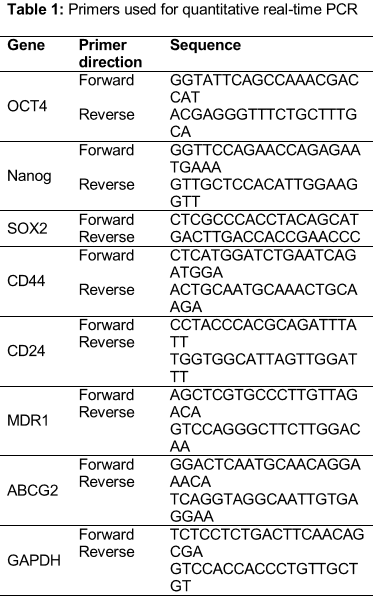
RNA extraction, RT-PCR and quantitative real-time PCR
Total RNA was extracted using TRIZOL Reagent (Invitrogen) and reverse transcribed with R-PCR Quick Master Mix (Toyoba) to produce cDNA. The primer sequences listed in were used for quantitative real-time PCR. Real-time PCR was performed using SYBR Green-based detection in LightCycler○R480 (Roche) according to the manufacturer’s instructions. GAPDH (glyceraldehyde-3' phosphate dehydrogenase) levels were used as normalization controls.
Chemoresistance assay
The MTT assay (Cell titer 96○R Aqueous One Solution Cell Proliferation Assay, Promega) was used to assess the rates of resistance to drugs. Briefly, breast cancer cells (2 × 10³/well) were seeded in 96-well plates. After 24 h in presence or absence of pectolinarigenin, the indicated concentration of doxorubicin (Ferrer Farma) was added and 72 h later, the MTT assay was performed using iMarkmicroplate Absorbance Reader (Bio-RAD, Richmond, CA) according to the manufacturer’s instructions.
Tumor growth in xenografts
MCF-7 Cells (5 × 10⁶) were injected subcutaneously in the right flank of each of 8 female 6-week old nude mice (Laboratory Animal Center, Xiamen University). All animals were maintained under controlled temperature, humidity, and 12 h light/dark cycles in the Xiamen University Laboratory Animal Center. The animals were fed standard rodent chow and allowed free access to water ad libitum. All experiments were carried out in accordance with NIH Guidelines for the Care and Use of Laboratory. Animal care and use was approved by the Ethics Committee of Faculty of Medicine-Xiamen University-China. When the tumors developed at 7 days, the mice were randomly distributed into two groups, of untreated or treated by i.p. injection every day with pectolinarigenin (20 mg/kg). Tumor volume (Tv) was measured every 3 days and calculated as in Eq 1.
Tv = (LW2)/2 ………………………….. (1)
where L and W are the length and breadth of the tumor, respectively.
Statistical analysis
The results are expressed as mean ± SEM. Statistical significance was determined by Student’s t-test or one-way or two-way analysis of variance (ANOVA) followed by Turkey’s test, as appropriate, using Graphpad Prism statistics software (Graphpad Software). p < 0.05 was considered statistically significant (*p < 0.05, **p < 0.01, ***p < 0.001).
Results
Pectolinarigenin inhibited BCSCs self-renewal properties
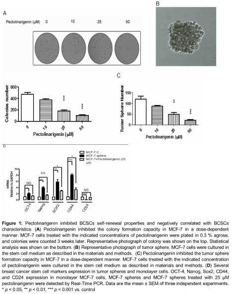
The isolated compound was identified by a combination of NMR and mass spectral data as reported previously [11]. Soft agar colony formation assay and tumor sphere assay have been widely used to identity stem cells in in vitro assays. The colony formation capacity in MCF-7 was firstly assayed under the treatment of pectolinarigenin. The results showed pectolinarigenin inhibited the colony formation capacity in MCF-7 cells in a dose dependent manner as shown in A. Even 25 μM concentration of pectolinarigenin could cause significant inhibition of colony formation in MCF-7 (61.23 %, p < 0.001, A).
Then the tumor sphere formation capacity was examined in MCF-7 under the treatment of pectolinarigenin. Tumor spheres of MCF-7 were cultured as described elsewhere [12]. Tumor spheres with a tight appearance were observed in serum free medium (B). Those results showed that the breast cancer cells sphere formation capacity was inhibited by pectolinarigenin in a dose dependent manner as shown in C, the inhibition efficiency of tumor sphere formation is 59.49 % when treated with 25 μM pectolinarigenin (p < 0.01, C).
Moreover, BCSCs marker expression in monolayer MCF-7 cells, MCF-7 spheres and MCF-7 spheres treated with 25 μM pectolinarigenin were compared by real-time PCR. The results showed that the spheres expressed much higher levels of BCSCs markers, such as OCT-4, Sox2, Nanog, CD44, than the monolayer cells. At the same time, the spheres expressed much lower levels of CD24 than the monolayer cells. The induction and repression of BCSCs markers were greatly reversed in the spheres treated with 25 μM pectolinarigenin compared with the untreated spheres (D).
Pectolinarigenin reduced breast cancer cell chemoresistance
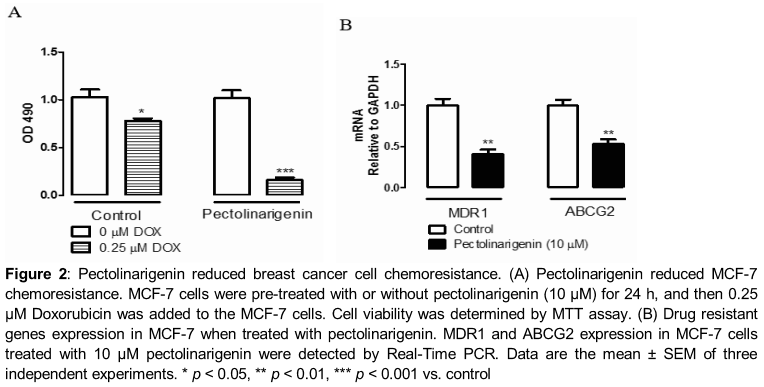
Studies in the past have suggested that chemoresistance is another characteristic of CSCs. Next studies were carried out to investigate whether pectolinarigenin would influence the chemoresistance of MCF-7 cells. For this purpose, MCF-7 cells were pre-treated with or without pectolinarigenin (10 μM) for 24 h and then 0.25 μM Doxorubicin was used to treat the cells. As showed in A, pectolinarigenin enhanced chemosensitivity to Doxorubicin. Moreover, we detected chemoresistant genes (MDR-1 and ABCG2) expression in MCF-7 when treated with pectolinarigenin (10 μM). As shown in B, MDR1 and ABCG2 expression were repressed under treatment with pectolinarigenin (10 μM). Especially, the inhibition efficiency of MDR1 and ABCG2 were about 59.29 % (p < 0.01, B) and 46.48 % (p < 0.01, B) when treated with pectolinarigenin as compared with the control, respectively.
Pectolinarigenin reduced tumorigenicity in vivo
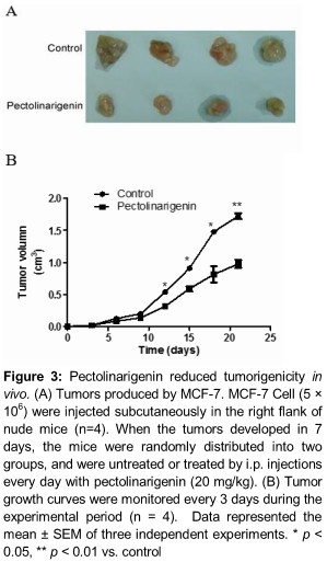
It has been reported that tumorigenicity in vivo correlates with the sphere formation ability of tumor cells in vitro. To test the effect of pectolinarigenin on the tumorigenicity of breast cancer cells, MCF-7 cells (5 × 10⁶) were inoculated into nude BALB/C mice. When the tumors developed in 7 days, the mice were randomly distributed into two groups, of untreated or treated with pectolinarigenin (20 mg/kg). The size and weight of xenograft tumors treated with pectolinarigenin were significantly smaller than control tumors (). These results thus demonstrated that pectolinarigenin efficiently attenuated the tumorigenicity of breast cancer cells in vivo.
Discussion
It is urgently required to develop an effective method of treatment of breast cancer, due to its high incidence and high recurrence rates in recent years. Hypothesis of cancer stem cells suggested that CSCs is the mainly responsible for causing relapse in cancer patients [2]. Bio-flavonoids extracted from fruits and vegetables have a variety of biological properties, including antibacterial, antioxidant, anti-inflammatory, antiviral, anticancer, cancer preventing activities [14,15]. Previously we have already demonstrated that the flavonoid pectolinarigenin inhibits the proliferation of breast cancer cell [11]. We speculated that inhibiting CSCs properties may be a potential mechanism of preventing carcinogenesis by pectolinarigenin. The tumor sphere assay has been used to identity breast cancer stem cells, as shown in D, CSCs markers (OCT4, Nanog, SOX2 and CD44) were greatly induced in our experimental system. The results demonstrated that pectolinarigenin inhibited breast cancer cell self-renewal capacity Consistent with the inhibitory effects, pectolinarigenin suppressed induction of BCSCs markers.
Another characteristic of CSCs is chemoresistance. MDR1 and ABCG2 play an important role in chemoresistance of breast cancer cells [15]. Our results showed that pectolinarigenin enhanced breast cancer cell chemosensitivity to doxorubicin. The chemosensitivity is associated with downregulation of MDR1 and ABCG2 (B). Pectolinarigenin inhibited MDA1 and ABCG2 mRNA expression, and at the same time, repressed tumor sphere formation. Our results are consistent with other reports that MDR1 and ABCG2 not only play a major role in multidrug resistance but also are characteristics of CSCs markers.
Conclusion
The findings of this study indicate that pectolinarigenin inhibits BCSCs properties in vitro, reduces breast cancer cell tumorigenicity in vivo, and provides an experimental basis for the improvement of breast cancer therapy.
Declarations
Acknowledgement
References
Archives


News Updates